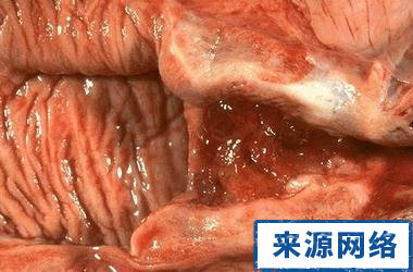
The function of the female reproductive organs is to reproduce offspring and perform sexual activities. There are many mysteries of the female reproductive organs. Let's take a look at the pictures of female vaginas and understand the common sense of women's physiology.
The vagina is a muscular tube composed of mucous membranes, muscularis and adventitia, rich in extensibility, connecting the uterus and external genitalia. It is the female sexual organ and the conduit for menstruation and delivery of the fetus. The normal pH of vaginal discharge is 3.8.
The vagina is often in a collapsed state where the anterior and posterior walls meet. The lower part of the vagina is narrow, and the lower end opens into the vestibule of the vagina through the vaginal orifice. There is a hymen attached to the virgin vaginal opening, which can be annular, half-moon-shaped, umbrella-shaped or sieve-shaped.
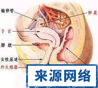
After the hymen is ruptured, there are hymen marks around the vaginal opening. The upper end of the vagina is broad and wraps around the cervix-vaginal part, forming an annular depression between the two, called the fornix of vagina, which can be divided into an anterior part, a posterior part and 2 lateral parts. The posterior part of the vaginal dome is the deepest, and is closely adjacent to the rectum-uterine depression, separated only by the vaginal wall and a layer of peritoneum. Clinically, it has great practical significance, such as the ability to drain the effusion in the depression through the posterior fornix.
Adjacent to the vagina: the bladder and urethra are located in the front, and the rectum is adjacent to the rear. The recto-uterine pit, cervix, and uterine orifice can be palpated clinically through the rectal wall. The lower part of the vagina passes through the urogenital diaphragm. Both the urethrovaginal sphincter in the diaphragm and the medial muscle fiber bundle of the levator ani muscle sphincter the vagina.


