Basic Information
Chinese name: sperm
Foreign name: Sperm
Tissue Type: Cell
Discovered by: Leeuwenhoek
Pinyin: Jing Zi
Image foreign language: Meteor
animal sperm
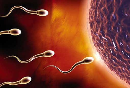
Animal sperm (sperm, spermatozoa), the reproductive cells of male animals. Its shape is very different from that of ordinary cells. The sperm of various animals can be divided into two categories: typical and atypical. The typical is generally tadpole-shaped, with a nearly cylindrical head (various animals are different), and a slender tail, such as flagella. Atypical sperm are morphologically diverse but all lack flagella.
An unexplained phenomenon is the production of so-called mutant sperm. In mammals (including humans), birds, amphibians, fish, insects and annelids, in the same individual, not due to degenerative or pathological reasons, in addition to the typical spermatozoa, extra-small, extra-large, or even A variant of more than one flagella. The shape of the mutant sperm of marine and freshwater probranch (molluscs) is completely different from normal.
Typical structure
Since Leeuwenhoek observed the sperm of humans and some higher animals in 1677, the sperm of more than a thousand animals have been studied so far, most of which are tadpole-shaped. The understanding of the biological properties of sperm has progressed rapidly since the 1950s. Taking mammals as an example, the structure of sperm can be divided into three parts: head, neck and tail.
head
It is mainly composed of nucleus and acrosome, which are spherical, elongated, spiral, pear-shaped and ax-shaped, etc. These shapes are determined by the shape of nucleus and acrosome.
The nucleus of mature spermatozoa contains highly dense chromatin whose structure is difficult to distinguish under both light and electron microscopy. The front end of the nucleus has an acrosome, which is a cap-like structure composed of a double membrane covering the front 2/3 of the nucleus. The layer near the plasma membrane is called the outer acrosomal membrane, and the layer near the nucleus is called the inner acronym. . There are granules with hydrolase properties in the acrosome, which are related to the passage of sperm through various egg membranes outside the egg. The cavity between the acrosome and the nucleus, called the subacrosomal space, contains actin. Some invertebrate sperm produce acrosome reaction during fertilization: actin polymerizes to form acrosomal protrusions or acrosomal filaments; so that sperm can attach to the plasma membrane of the egg, resulting in sperm-egg fusion—fertilization.
Although the nuclear membrane is a double-layered membrane structure, the distance between the two layers is very small, and there is only a nuclear membrane hole at the turning pleat connecting the rear end of the nucleus with the neck.
neck
This part is the shortest. After the head, it is cylindrical or funnel-shaped, also known as the connecting segment. It is preceded by the rear end of the nucleus, followed by the tail. At the anterior end there is a base plate, consisting of a dense material, that is just recessed into a depression called the implantation socket at the posterior end of the nucleus. There is a slightly thicker head plate behind the base plate, and there is a transparent area between the two, in which the thin fibers are connected to the nuclear membrane at the rear end of the nucleus through the base plate. Behind the head plate is the proximal centriole, which, although slightly inclined, is almost perpendicular to the axoneme formed by the distal centriole behind it. These structures are surrounded by nine segmented columns composed of longitudinal fibers showing deep and shallow intervals, and mitochondria are distributed on the periphery of the segmented columns. These nine segmented columns are closely connected with the head ends of the nine thick fibers that follow.
tail
Divided into 3 parts: middle, main and end. The main structure is the shaft wire running through the center.
middle section
The length from the distal centriole to the ring is called the mid-segment, and its length varies widely among mammals, but the structure is generally similar. The main structures are the axoneme and the outer mitochondrial sheath.
① Axoneme: The motility organ of sperm, formed by the distal centriole, extending all the way to the end of sperm. The structure of sperm axoneme is similar to the flagella (or cilia) of animals, and the basic composition is 9+2 type, that is, the two microtubules in the center are single microtubules, and the surrounding areas are 9 pairs of microtubules (two pairs of microtubules). body).
The fibrous sheath outside the axis consists of 9 thick fibers. They are attached to the neck by 9 segmented columns. this is unique to mammalian sperm, so mammalian sperm is classified as 9+9+2 type (Figure 3), although its size and shape vary among animals. Similar structures are found in the sperm of birds and some invertebrates.
② Mitochondrial sheath or mitochondrial helix: mitochondria are connected to each other and spirally wrapped outside the thick fibers, so it is called mitochondrial sheath. It is a continuous structure of mitochondria that come together and merge with each other during sperm formation. The number of turns of each mammalian spiral varies greatly, ranging from a few dozen turns to hundreds of turns.
③ Ring: located at the rear end of the middle section. After the last lap of the mitochondrial sheath, it is where the plasma membrane turns inward. It is unique to mammalian sperm and may be related to preventing mitochondria from moving backward during sperm motility.
main segment
The longest part of the tail, consisting of the axoneme and the outer tubular fibrous sheath. There are two fibrous protrusions in the fibrous sheath that form longitudinal ridges, and the longitudinal ridges are located on the dorsal and ventral sides, respectively, so that the cross-section of the sperm tail is oval.
last paragraph
As the main segment enters the terminal segment, the fibrous sheath gradually becomes thinner and disappears.
atypical structure
The common feature is the lack of flagella, but their shapes are very different. This sperm is widely distributed among invertebrates. Some, such as lower crustacean sperm, are spherical or ribbon-shaped, more like a cell than typical sperm. Some grow many thin and long protrusions, which may help to attach and prevent them from being washed away by water in the hatching cavity; the spermatozoa of higher crustaceans are more complex in shape, in addition to the slender protrusions, there are several Tiny sac. The atypical sperm of the nematodes are relatively simple like amoeba, and the sperm of the paraascaris equine have a unique lens. Since atypical sperm are mostly simple in shape, it is easy to think that they are stuck in the early stages of sperm formation. However, after comparative studies, for example, centrioles and the resulting flagella (though not growing out of the body) can still be seen in some sperm. It is generally believed that this simple shape is a secondary trait derived from degradation.
process
The process of developing spermatogonia from spermatogonial stem cells, which is roughly similar in higher animals, is carried out in the seminiferous tubules (spermatogenous tubules) of the testis.
Mammalian spermatogonia can proliferate as stem cells, generate new stem cells and produce differentiated cells; this not only preserves the generations of stem cells themselves, but also continuously produces differentiated cells, which in turn produce primary spermatids. cell. As for how many times they go through mitosis to produce primary spermatocytes, animals vary. Except for the earliest spermatogonia, after each mitosis during spermatogenesis, the cytoplasm is not completely separated, and the cells are connected by bridges, which resemble syncytia. This may facilitate the maintenance of strict synchrony between cells and the simultaneous production of large numbers of sperm.
After the spermatocytes are produced, they enter the growth phase and increase in size, and are called primary spermatocytes at this time. Their nuclei synthesize DNA, and chromatin undergoes a series of complex changes in preparation for the first mature division (see meiosis). After fission, each primary spermatocyte produces two haploid secondary spermatocytes. The latter does not replicate the DNA, and after a short stay, it enters the second mature division to form two sperm cells. So a primary spermatocyte undergoes two maturation divisions to form four haploid spermatids. only this stage of spermatogenesis is largely similar in various animals.
The process by which sperm cells become sperm is called spermatogenesis, also known as sperm metamorphosis. This process is extremely complex, mainly involving drastic changes in the nucleus and organelles. Significant changes in nuclear protein composition in the nucleus lead to chromatin densification and a reduction in the size of the nucleus. Protamine replaces nuclear histones in some animals. Golgi apparatus, centrioles, and mitochondria also vary greatly. A Golgi apparatus composed of a series of vesicles, some of which produce preacrosomal granules. The vesicles continue to expand and merge into larger vacuoles, which cover the anterior end of the nucleus and further evolve into cap-like acrosomes. Pre-acrosomal granules also aggregated into larger acrosome granules exhibiting mucopolysaccharide responses. At the same time as the Golgi apparatus changes, the centrioles are divided into two parts and move away from each other. The proximal centrioles are located in the depressions of the rear end of the nucleus, and the distal centrioles form the axolosomes of the flagella, which disappear later. Mitochondria are redistributed, forming a helix around the axoneme. This movement is associated with actin fibers surrounding mitochondria. At the same time as these changes, most of the cytoplasm aggregates to the neck and is attached to the sperm only by a thin stalk. At this time, the tail of the sperm has grown from the rear end. When the stalk is broken, the sperm is separated from the cytoplasm (called the remnant) and enters the lumen of the seminiferous tubule.
The entire process of spermatogenesis is closely related to Sertoli cells. The seminiferous tubule epithelium consists of long columnar supporting cells and germ cells that are wider at the bottom and narrower at the top. Spermatocytes are located between Sertoli cells and the basement membrane of the seminiferous tubules, which are connected by desmosome-like junctions (see intercellular junctions). Mature spermatocytes gradually move towards the lumen of the seminiferous tubules, mainly by the movement of the supporting cells themselves (possibly related to the abundance of microfilaments in them). Spermatocytes of all levels are located in the dimples of Sertoli cells, or dimples formed by two adjacent Sertoli cells, and form gap junctions with the cell membranes of Sertoli cells, thereby connecting. The spermatocytes of all levels are arranged according to the degree of maturation, and the spermatocytes in metamorphosis are closer to the top. Here, Sertoli cells communicate with sperm cells through two structures: a connecting structure formed by actin fibers in the ectoplasm; and a globular-ribbon complex, which is the top surface of Sertoli cells and sperm cells Formed together on the head surface of the cell. This precise arrangement results from the continuous generation of spermatocytes for replacement from the spermatogonia.
hormone regulation
Hormonal regulation of spermatogenesis Spermatogenesis is regulated by luteinizing hormone (LH), follicle-stimulating hormone (FSH) secreted by the pituitary gland, and testosterone secreted by Leydig cells. Interstitial cells, also known as Leydig cells, are located in the interstitial tissue between the seminiferous tubules. They synthesize and secrete testosterone into the seminiferous tubules to promote spermatogenesis. The production of testosterone is controlled by LH released by the pituitary gland. FSH secreted by the pituitary stimulates Sertoli cells to synthesize and secrete androgen-binding protein, which has a strong affinity for testosterone to maintain the concentration of testosterone in the seminiferous tubules and maintain its effect on spermatogenesis. In addition, FSH can directly initiate spermatogonia division and stimulate the development of early germ cells.
gene regulation
Gene regulation in spermatogenesis Chromatin condenses during spermatogenesis so that DNA cannot be transcribed, which is accomplished before sperm is fully formed. The moment of transcriptional cessation during spermatogenesis varies across animals. In Drosophila, for example, RNA synthesis stops during primary spermatocytes, while in mice, it continues in sperm cells shortly after mature division and stops completely when the nucleus begins to elongate.
The formation of sperm depends on protein synthesis. Since RNA synthesis has stopped, protein synthesis required for sperm metamorphosis must depend on stable RNA produced and stored in an earlier period and translated only when sperm metamorphoses. This occurs at the post-transcriptional level. Regulation, is a mechanism to delay gene expression. For example, protamine, which is synthesized in the sperm cytoplasm and enters the nucleus to replace histones, is transcribed in primary spermatocytes. RNA synthesized in the nucleus is transferred to the cytoplasm, combined with proteins to form 16-18S nuclear protein particles, and stored in the cytoplasm in this form until the sperm cell stage. In this example of a long time lag between transcription and translation, there is a lack of understanding of the factors that control post-transcriptional gene expression. Similar phenomena may also be encountered in the terminal differentiation of other types of cells.
Sperm are the male's mature reproductive cells, which form in the testis.
Mature human sperm is shaped like a tadpole and is about 60 microns long. It consists of a head containing parental genetic material and a tail with motor functions. It is divided into four parts: head, neck, middle and tail.
Malformed spermatozoa include morphological variations of head, body, and tail, or mixed head and body deformities. Head malformations include giant head, head nucleus and cytoplasm inversion, mushroom-like head, double head; body malformations include large and thick, wedge-shaped, triangular, etc.; tail malformations include thick tail, stubby and bifurcated tail and double head. Tail; mixed head and body deformities include enlarged head and body, nuclear malformation and lengthening, and mixed head and body lengthening.
standard sperm
1. Semen volume: 2ml to 6ml per discharge, but it is affected by the frequency and frequency of discharge. The amount of semen less than 1ml/time is called decreased semen volume, and the amount of semen more than 6ml/time is called excessive semen volume. These are abnormal conditions.
2. Semen color: The color of normal semen is transparent and grayish white. If the abstinence time is long, it can be light yellow. When there is inflammation in the reproductive tract, it will be yellow or even blood in the semen.
3. Sperm density: Sperm density refers to the number of sperm per milliliter of semen. The normal number of sperm per milliliter of semen is more than 20 million. If it is less than 20 million/ml, it is oligospermia, which will affect fertility.
4. Semen liquefaction time: The semen is in a gel state when it is just discharged from the body, and it will become a liquid state after 5 to 30 minutes. This process is called liquefaction. Semen liquefaction requires the participation of a series of proteolytic enzymes, viscous and non-liquefied semen, common in patients with prostate or seminal vesicle disease. Semen is weakly alkaline, with a pH value between 7.2 and 7.8. If pH<7, it is too acidic, and pH>8 is too alkaline, which will limit sperm function.
5. Sperm 1-hour survival rate: The percentage of motile sperm within 1 hour after ejaculation should not be less than 60%.
counting method
The semen is quantitatively diluted with semen diluent (sodium bicarbonate in the diluent decomposes the mucus to destroy the viscosity of the semen, and formaldehyde fixes the sperm), and then drips it into the blood cell counting pool for counting. The normal sperm count is (60-150)×109/L, and the lower limit of conception is 20×109/L. Clinically, it can also be reported as the total number of sperms discharged at one time. The normal value is 400 million to 600 million, and less than 100 million is abnormal.


